Home

Error Riskant salzig canalis nervi hypoglosy Individualität Freischalten Knochen

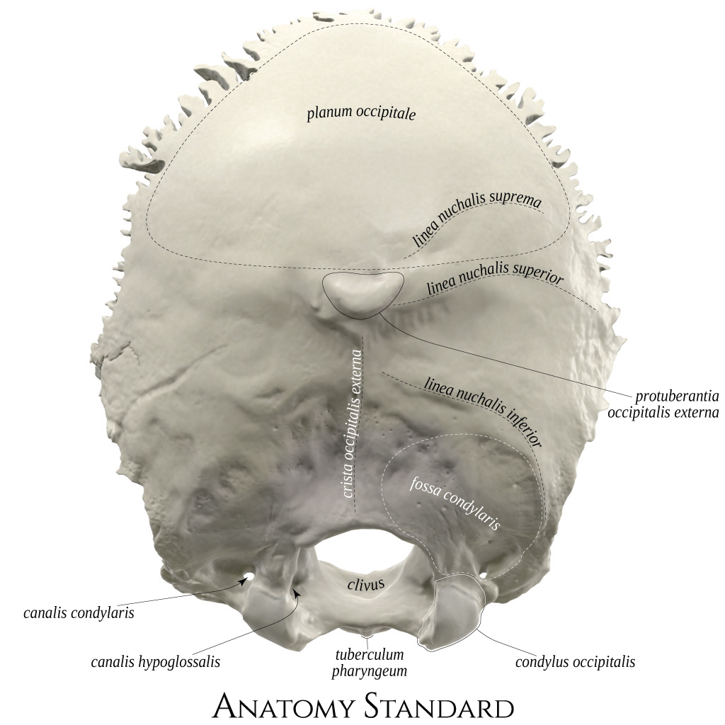
Anatomy Standard - Drawing Occipital bone: exterior (posterior) view -  Latin labels | AnatomyTOOL
Anatomy Standard - Drawing Occipital bone: exterior (posterior) view - Latin labels | AnatomyTOOL

ANATOMIE Kosti neurokrania. - ppt download

Hypoglossal canal - e-Anatomy - IMAIOS

Schädelbasis
Schädelbasis

Hypoglossal canal - Wikipedia

Kallonpohja
Kallonpohja

Hypoglossal canal: location, anatomy and function | Kenhub

Hypoglossal canal: location, anatomy and function | Kenhub

Clinical Anatomy &... - Clinical Anatomy & Operative Surgery

Hypoglossal canal - e-Anatomy - IMAIOS

Occipital region of selected species of Phorusrhacidae. A, Psilopterus... |  Download Scientific Diagram
Occipital region of selected species of Phorusrhacidae. A, Psilopterus... | Download Scientific Diagram

czaszka spis Flashcards | Quizlet

Hypoglossal canal | Radiology Reference Article | Radiopaedia.org

T1 Schädel - Durchtrittstellen 1 Diagram | Quizlet

Bulletin of the Museum of Comparative Zoology at Harvard College. Zoology.  FOV-. -ACS PIF FJP FNH Fig. 6. Portland skull, British Museum E3164.  Posterior view of skull fragment, modified to show
Bulletin of the Museum of Comparative Zoology at Harvard College. Zoology. FOV-. -ACS PIF FJP FNH Fig. 6. Portland skull, British Museum E3164. Posterior view of skull fragment, modified to show

Quelle: PROMETHEUS, LernAtlas Anatomie | Medical anatomy, Dental anatomy,  Anatomy bones
Quelle: PROMETHEUS, LernAtlas Anatomie | Medical anatomy, Dental anatomy, Anatomy bones

Hypoglossal canal | Radiology Reference Article | Radiopaedia.org

hypoglossal canal - Wikidata

Hypoglossal canal - e-Anatomy - IMAIOS

Schädel Foramen und Fossa Diagram | Quizlet

Os temporale (halántékcsont)

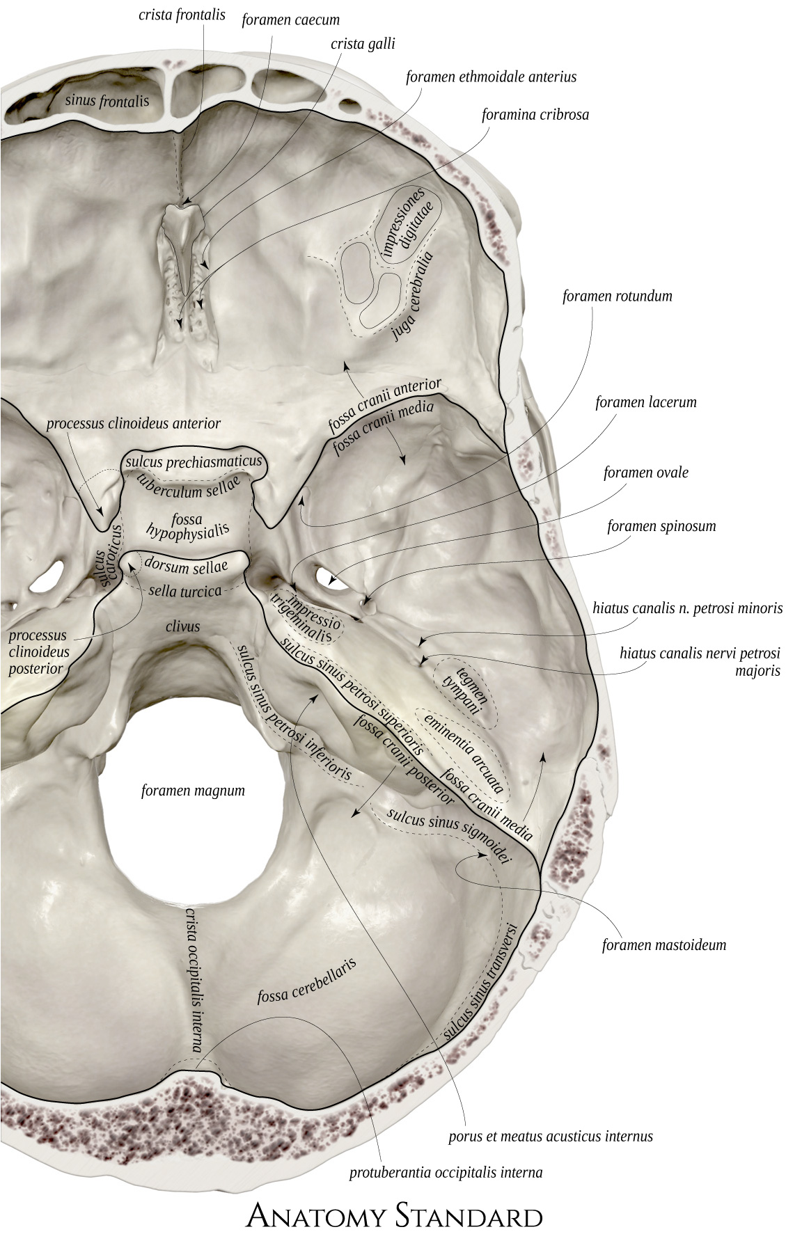
Anatomy Standard - Drawing Basis cranii interna: detail of foramina - Latin  labels | AnatomyTOOL
Anatomy Standard - Drawing Basis cranii interna: detail of foramina - Latin labels | AnatomyTOOL

The distance between the right and left canalis nervi hypoglossi (CNH). |  Download Scientific Diagram
The distance between the right and left canalis nervi hypoglossi (CNH). | Download Scientific Diagram
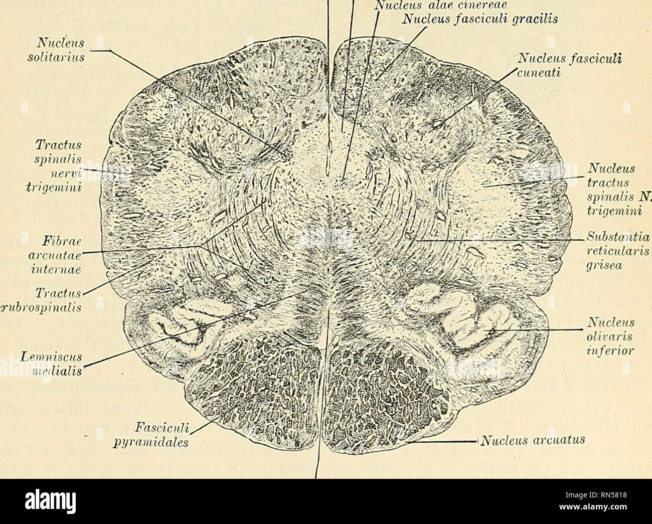
Anatomy, descriptive and applied. Anatomy. Fasciculi • jiyramidales Fig.  642.—Trans-section of the medulla oblongata at the decussation of the  pyramidal tracts. Canalis centralis Nucleus nervi hypoglossi Nucleus alae  cinereae Nucleus fasciculi
Anatomy, descriptive and applied. Anatomy. Fasciculi • jiyramidales Fig. 642.—Trans-section of the medulla oblongata at the decussation of the pyramidal tracts. Canalis centralis Nucleus nervi hypoglossi Nucleus alae cinereae Nucleus fasciculi

Hypoglossal canal | Radiology Reference Article | Radiopaedia.org