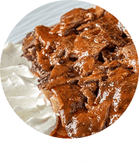
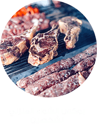
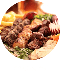
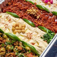
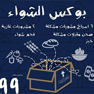
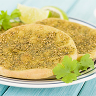
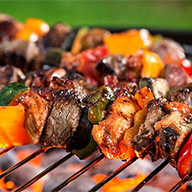
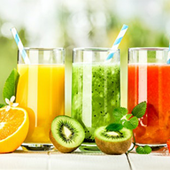
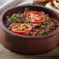
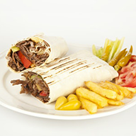
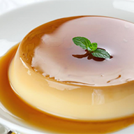
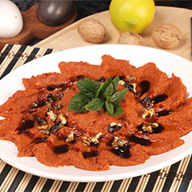
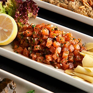
![The left image is an MRI GRE sequence, showing the hemosiderin ring... | Download Scientific Diagram The left image is an MRI GRE sequence, showing the hemosiderin ring... | Download Scientific Diagram](https://www.researchgate.net/publication/326575726/figure/fig7/AS:651913146138638@1532439614544/The-left-image-is-an-MRI-GRE-sequence-showing-the-hemosiderin-ring-arrow-and-the-right.png)
The left image is an MRI GRE sequence, showing the hemosiderin ring... | Download Scientific Diagram
![Hemosiderin detection in human CCM tissues. (A) TA/SRS image and (B)... | Download Scientific Diagram Hemosiderin detection in human CCM tissues. (A) TA/SRS image and (B)... | Download Scientific Diagram](https://www.researchgate.net/publication/326441476/figure/fig5/AS:649351139442697@1531828784673/Hemosiderin-detection-in-human-CCM-tissues-A-TA-SRS-image-and-B-Perls-staining-of.png)
Hemosiderin detection in human CCM tissues. (A) TA/SRS image and (B)... | Download Scientific Diagram
![Neurology Review on Twitter: "♦️They have characteristic pattern on T2-weighted images, with a central nidus of irregular bright signal intensity mixed with mottled hypointensity (“popcorn” pattern), surrounded by a peripheral hypointense ring Neurology Review on Twitter: "♦️They have characteristic pattern on T2-weighted images, with a central nidus of irregular bright signal intensity mixed with mottled hypointensity (“popcorn” pattern), surrounded by a peripheral hypointense ring](https://pbs.twimg.com/media/EGi-69zXkAQ72XI.jpg)
Neurology Review on Twitter: "♦️They have characteristic pattern on T2-weighted images, with a central nidus of irregular bright signal intensity mixed with mottled hypointensity (“popcorn” pattern), surrounded by a peripheral hypointense ring
![Frontiers | 1.5 Tesla Magnetic Resonance Imaging Features of Canine Intracranial Intra-axial Hematomas Frontiers | 1.5 Tesla Magnetic Resonance Imaging Features of Canine Intracranial Intra-axial Hematomas](https://www.frontiersin.org/files/Articles/778320/fvets-08-778320-HTML/image_m/fvets-08-778320-g001.jpg)
Frontiers | 1.5 Tesla Magnetic Resonance Imaging Features of Canine Intracranial Intra-axial Hematomas
![Neurovascular Lesions in Pediatric Epilepsy - J. Kassiri, T. Rajapakse, M. Wheatley, D. B. Sinclair, 2019 Neurovascular Lesions in Pediatric Epilepsy - J. Kassiri, T. Rajapakse, M. Wheatley, D. B. Sinclair, 2019](https://journals.sagepub.com/cms/10.1177/0883073819838445/asset/images/large/10.1177_0883073819838445-fig1.jpeg)